Applications
Biophysics
- Application note
- No. BPS001-IFDS-20240419-001
Dark-field spectroscopy of plasmon-enhanced scattering and fluorescence from polymer-coated silver nanoparticles
- Keywords
- Plasmon biosensors
- Metal nanoparticles
- Localized surface plasmon resonance (LSPR)
- Dark-field microscopy
- Dark-field spectroscopy
- InFocus λ DS
In the R&D of plasmon biosensors utilizing metal nanoparticles, dark-field microscopy for nanostructure observation and spectroscopy for optical response investigation are indispensable. Here, we present an example of dark-field spectroscopic analysis focusing on surface plasmon-enhanced scattering and fluorescence from Ag nanoparticles, utilizing a dark-field spectroscopic imaging microscope.
Published Online: Apr. 19th, 2024
Download PDFIntroduction
Fluorescence spectroscopy using metal nanoparticles has attracted attention as a highly sensitive optical biosensing technique due to the field enhancement effect via localized surface plasmon resonance (LSPR), which enhances the fluorescence intensity of labeled molecules.
The resonance wavelength of LSPR varies depending on factors such as the size and shape of the metal nanostructures, the type of metal (e.g., gold or silver), and the refractive index of surrounding media and adsorbed substances near the metal surface. In LSPR biosensors, ligand molecules binding to the target substance are coated on the metal surface, and their binding is detected as spectral changes.
To advance the application of metal nanoparticles in biosensing, evaluating the optical properties of individual particles is crucial, necessitating dark-field observation at the nanoscale and spectroscopic analysis of scattered light and fluorescence. Here, we utilized the ScienceEdge InFocus λ DS, a dark-field spectroscopic imaging microscope, to perform dark-field spectroscopy of plasmon-enhanced scattered light and fluorescence using polymer-coated silver nanoparticles.
Sample preparation
When fluorescent molecules are in proximity to a metal surface, plasmonically enhanced fluorescence increases significantly, but simultaneous quenching of fluorescence occurs due to energy transfer
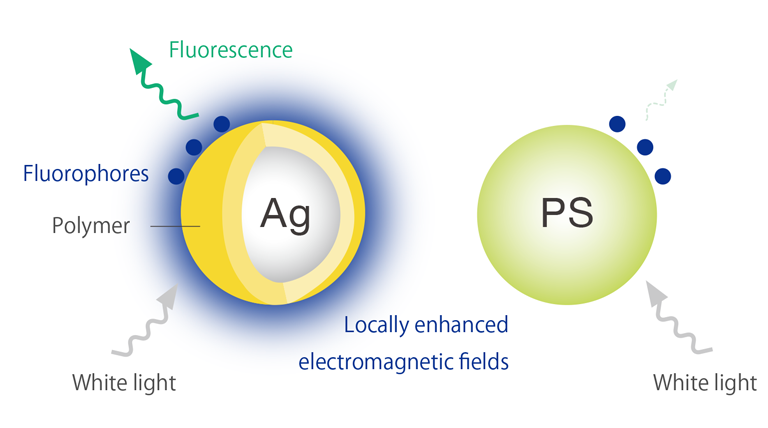
Figure 1. Fluorophores are excited by the locally enhanced electromagnetic fields. In the case of PS nanoparticles, fluorophores are not excited because no electric field enhancement occurs.
1
2
from the fluorophore to the metal. To achieve high field enhancement while suppressing this quenching effect, a method of forming a few nanometers thick spacer film on the surface of metal nanoparticles is known.
Here, we measured 80 nm diameter silver nanoparticles coated with a 4 nm thick functional polymer using the InFocus λ DS, observing the dark-field images of isolated silver nanoparticles and performing dark-field spectroscopy of enhanced scattered light. Additionally, we conducted surface plasmon-field enhanced fluorescence spectroscopy (SPFS) for polymer-coated silver nanoparticles modified with fluorescent dye molecules (Alexa Fluor: λex = 430 nm, λem = 540 nm).
Dark-Field Spectroscopic Measurement
The configuration of the InFocus λ DS is illustrated in Figure 2. Using a ring diaphragm and a ring mirror, ring-shaped white LED light is obliquely illuminated to avoid direct entry into the observation optical system. To perform fast hyperspectral imaging under dark-field microscopy, the slit direction of the spectrograph is utilized for spatial information, enabling simultaneous spectral measurements along the X-axis of the specified measurement area (line detection). By scanning the detection position in the Y-axis direction using a galvanometer scanner, dark-field spectroscopic imaging can be executed rapidly. With a 256 x 1024 pixel cooled CCD, a large amount of data can be acquired both spatially and spectrally.
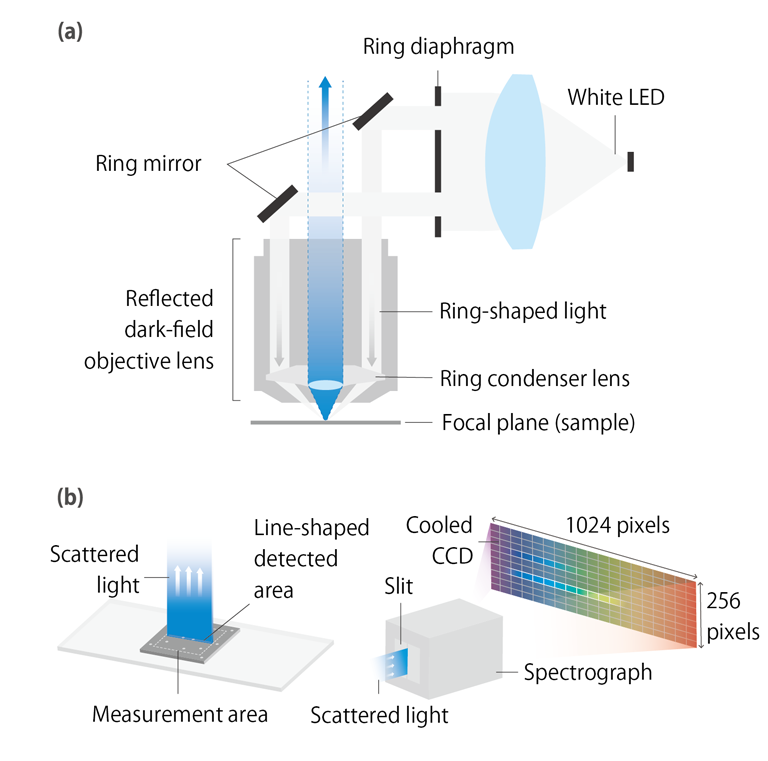
Figure 2(a). Optical system configuration of reflected-type dark-field microscopy. (b). Line detection in dark-field spectroscopy. 256 points on the X-axis are detected as spectra simultaneously.
2
3
Results
Figure 3(a) shows a dark-field microscope image of polymer-coated silver nanoparticles, revealing the distribution of individual particles with enhanced blue scattered light due to LSPR. Figure 3(b) presents the dark-field scattered spectrum of the designated area in Figure 3(a), demonstrating a distinct LSPR peak at approximately 460 nm. This wavelength overlaps with the absorption wavelength of the fluorescent dye, enabling excitation of the dye molecules by plasmons.
Figure 4 displays the emission spectrum from fluorescent dye molecules excited by plasmons on polymer-coated silver nanoparticles, with a peak at approximately 540 nm. To estimate the fluorescence enhancement factor, we measured the fluorescence spectrum of a single dye-functionalized polystyrene (PS) nanoparticle with a diameter of 100 nm. Since PS nanoparticles do not have a resonance wavelength in the visible range, fluorescence from PS nanoparticles is too weak to be detected (black line). Assuming the fluorescence intensity from these PS nanoparticles as the noise level of the spectrum, the fluorescence intensity ratio between polymer-coated silver nanoparticles and PS nanoparticles is estimated to be at least 20-fold. Considering the difference in surface area between polymer-coated silver nanoparticles and PS nanoparticles, the fluorescence enhancement factor of polymer-coated silver nanoparticles is estimated to be at least 26-fold.
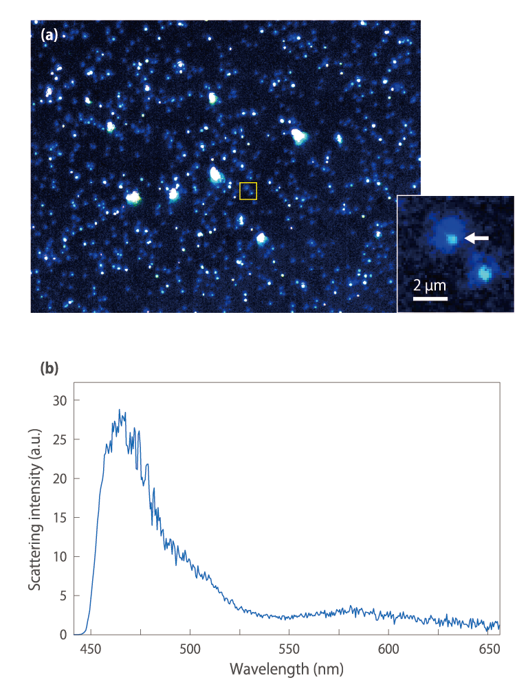
Figure 3 (a). Dark-field microscopic image of polymer-coated silver nanoparticles. The distribution of individual particles can be observed, and it can be confirmed that blue scattered light is enhanced by LSPR. (b). Dark-field scattering spectrum of the particle indicated by the white arrow in the enlarged view of Figure 3(a). It shows a peak near 460 nm, which is the characteristic plasmon scattered light from silver nanoparticles.
3
4
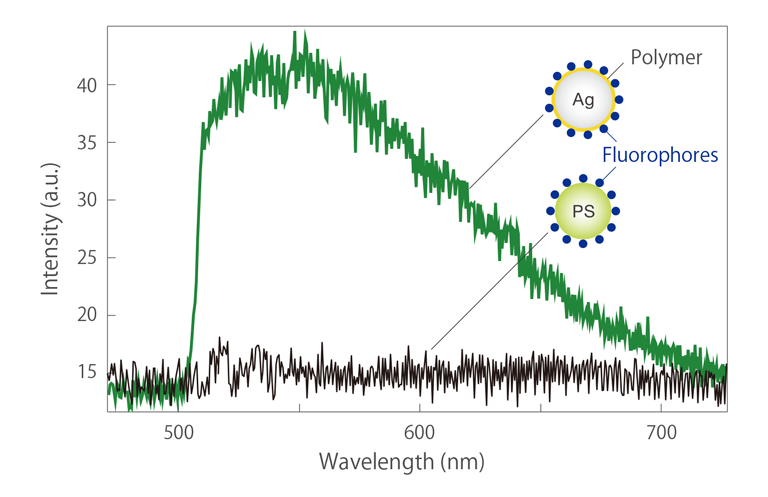
Figure 4. The emission spectrum (green) originating from fluorescent dye excited by plasmon on polymer-coated silver nanoparticles. The spectrum measured from PS nanoparticles modified with the same dye (black) shows only noise components, with no fluorescence observed.
ScienceEdge's InFocus λ DS combines dark-field microscopy with fast spectroscopic imaging capability, making it a powerful analysis tool for the further development of plasmon biosensors.
Acknowledgments
We deeply appreciate Dr. Taka-aki Yano of Tokushima University for providing the measurement data and valuable comments on this application note.
References
Kato et al., ACS Omega, 2022, 7, 4286−4292.
